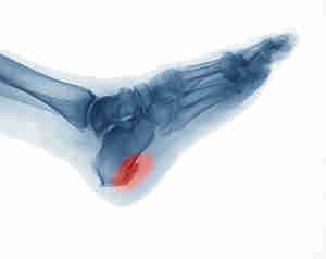
Plantar Fasciitis, Heel Spurs and Cortisone Injections

What could be causing my sore heel?

Plantar fascia is a thick fibrous band of connective tissue originating on the bottom surface of the heel bone and extending along the sole of the foot towards the toes. It acts as a passive limitation to the over-flattening of your arch.

Plantar heel pain accounts for approximately 11-15% of all foot symptoms requiring medical attention in adults and for 8-10% of all running-related injuries. (Rasenberg et al, 2018)

What is it?

Plantar fasciopathy (previously known as ‘Plantar Fasciitis’) is an overuse condition at the point where the band of fascia attaches to the calcaneus (heel bone). People typically first notice pain under their heel or in their foot arch in the morning or after resting.

Why is it called fasciopathy now instead of plantar fasciitis?

The term “itis” means inflammation. On ultrasounds and MRI, thickening and degenerative tissue findings of the fascia are more commonly seen than inflammatory changes. “opathy” Simply means pathology (or problem) of the tendon so “fasciopathy” has become the more correct term.

Who is affected?

Heel pain commonly affects very physically active people (e.g. runners) or people with high amounts of standing occupational work (who may also have a high BMI). 1 in 10 people will suffer from plantar fasciopathy in their lifetime.

Are Heel Spur’s causing my pain?

A heel spur is a calcium deposit causing a bony protrusion on the underside of the heel bone. They are very common in people WITHOUT heel pain and there is a very poor correlation between spurs and pain. If you have been diagnosed with a heel spur it is unlikely to be causing your pain. It is very likely to be plantar fasciopathy due to its location (attaching under the heel).

How do we diagnose it?

The most important part of the diagnosis is the history. Patient’s will report a gradual onset of heel pain that it worse in the mornings when getting out of bed (“first-step pain”) and then warms up with activity. It may then return as an ache post-activity of after sitting long periods.

There is pain or tenderness along the medial/dorsal aspect (inside/underneath) of the heel bone, which may extend along the medial and central components of the plantar fascia. Stretching the plantar fascia via lifting the big toe up (during the windlass test) may reproduce pain and may assist with palpation of the plantar fascia.

Do I need to get a scan?

X-rays and MRI’s are not indicated if there is a classic history and examination findings.

What are some other possible causes of heel pain?

Bruised heel (Fat pad contusion):

A bruised heel is an injury to the fat pad that protects the heel bone. It can occur acutely and chronically due to poor heel cushioning or repetitive stops, starts and changes in direction. Pain is felt laterally in the heel during weight-bearing activities due to the pattern of heel strike. Tenderness is felt in the posterolateral heel region. This activity helps with differentiating fat pad contusion with plantar fasciopathy.

Calcaneal stress fracture:

Occurs in runners, ballet dancers and jumping athletes. History of insidious onset of heel pain that is aggravated by weight-bearing activities, especially running. Presenting with localised tenderness over the medial or lateral aspects of the heel bone. Pain is reproduced by squeezing the posterior aspect of the calcaneus (heel bone) from both sides simultaneously.

Nerve pain:

This condition presents with rear foot pain caused by entrapment of the first branch of the lateral plantar nerve. Pain radiates to the medial inferior aspect of the heel proximally into the medial ankle region. Patients do not normally complain of numbness in the heel or foot. Often confused with plantar fasciopathy, the two conditions were shown to coexist in 52% of patients with this nerve entrapment. There is maximal tenderness at the plantar medial heel just proximal to the plantar fascia.

Midfoot pain:

Acute midfoot pain results from a sprain of the mid tarsal joint or plantar fascia. Gradual pain is a sign of overuse injury such as extensor tendinopathy, tibialis posterior tendinopathy or navicular stress fracture (pain poorly localised).

Other:

Blood tests may be warranted to rule out inflammatory arthritis, which can cause symptoms in the heels (although frequently symptoms will be more widespread). Plain radiographs (lateral view) are used to investigate the presence of a ‘calcaneal spur’. MRI is useful for identifying stress fractures of the calcaneus and other soft tissue pathologies; soft tissue tumours, medial tendinopathy and plantar fat pad pathology.

How do I treat plantar fasciopathy?

Research shows the best long term relief of heel pain is strength training. (Almubarak et al., 2012)

The 2 best strength exercises:

CLICK HERE TO SEE THE VIDEO!

1. Intrinsic foot muscles:

Begin sitting with your feet flat on the ground. Raise your arch by curling the big toe towards your heel, without curling your toes or lifting your heel.

2. Calf raises:

High load strength training consisting of heel-raises with a tight band around your ankles. Progress by performing on one leg at a time or by placing a towel under the toes.

What can I do for short-term relief?

The best short term relief of heel pain includes stretching and corticosteroid injections. (Babatunde et al., 2018)

The 3 best Stretching/flexibility exercises:

1. Big toe extension with ankle dorsiflexion:

Holding the big toe into slight extension and bringing the foot back into dorsiflexion.

2. Calf stretch:

Standing with your hands leaning against a wall for support and place the affected leg behind. Keep the leg straight and slight lean forward at the ankle joint. Feeling the gentle stretch in the calf muscle.

3. Self-massage with a golf ball:

Place a golf ball under the bottom of your foot and gently roll the ball back and forth.

Do Corticosteroid injections work?

Corticosteroid injections, alone or in combination with exercise were ranked most likely to be effective for management of short-term pain and function in the ‘Comparative effectiveness of treatment options for plantar heel pain’ study. However, in the long term, only exercise was beneficial.

What doesn’t work?

  • Heat/ice pack
  • Massage
  • Orthotics
  • Interferential
  • Ultrasound
  • Acupuncture

These options provide short term pain relief of up to 2-3 hours post treatment and are not as effective as exercise therapy. Interventions such as exercise (strength and flexibility) provide excellent long term benefits and pain relief solutions for plantar fasciopathy.

Foot orthoses vs Stretching

A systematic review and meta-analysis in the ‘Efficacy of foot orthoses for the treatment of plantar heel pain,’ explores the effect of different foot orthoses versus stretching exercises (Achilles and plantar fascia). The study showed that foot orthosis interventions are not superior in improving pain, function or self-reported recovery when compared with other conservative interventions in patients with plantar heel pain.

Orthotics vs Exercise therapy

The systematic review of ‘Exercise Therapy for Plantar Heel Pain,’ discussed the effect of exercise therapy versus the combination of customised orthotic devices and exercise. The trial concluded that both exercise therapy and combination of exercise with a customised orthotic device reduced the overall foot pain when compared with baseline results. However, the exercise therapy alone group was more effective in reducing overall pain than the combination group in those who stood for 8 hours or more daily.

The findings of this review support the use of exercise therapy following acute and chronic plantar heel pain over control/sham therapy, repetitive shock wave therapy, NSAIDs and orthotic devices.

References

Almubarak, A., & Foster, N. (2012). Exercise Therapy for Plantar Heel Pain: A Systematic Review[Ebook].

Babatunde, O., Legha, A., Littlewood, C., Chesterton, L., Thomas, M., & Menz, H. et al. (2018). Comparative effectiveness of treatment options for plantar heel pain: a systematic review with network meta-analysis. British Journal Of Sports Medicine, 53(3), 182-194. doi: 10.1136/bjsports-2017-098998

Brukner, P., Clarsen, B., Cook, J., Cools, A., Crossley, K., & Hutchinson, M. et al. (2017). Clinical Sports Medicine (5th ed.).

Rasenberg, N., Riel, H., Rathleff, M., Bierma-Zeinstra, S., & van Middelkoop, M. (2018). Efficacy of foot orthoses for the treatment of plantar heel pain: a systematic review and meta-analysis. British Journal Of Sports Medicine, 52(16), 1040-1046. doi: 10.1136/bjsports-2017-097892



Tennis Elbow – Latest Evidence

What is Tennis Elbow?

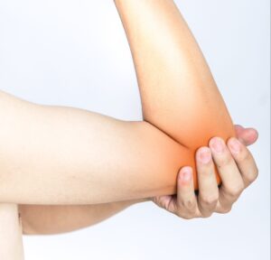
Tennis elbow is defined as a cause of pain and tenderness in the outer part of the elbow (lateral epicondyle) where the tendons of the forearm muscles attach.

It is an overuse injury from repetitive or forceful/explosive movements involving eccentric motion and/or in which the wrist frequently deviates from a neutral position. This can be from training errors, inadequate equipment or poor environmental conditions.

Who can get Tennis Elbow?

Tennis elbow can affect anyone, however is more common in people between 30 and 60 years of age. It appears to be more severe and of longer duration in females. The most commonly affected arm is the dominant arm. It is commonly seen in office workers (repetitive typing) or manual labour workers (carpenters etc).

Signs and Symptoms of Tennis Elbow?

Pain and tenderness over the elbow bone (lateral epicondyle)

Pain with gripping, twisting, lifting.

Some cases may have nerve involvement – nerve pain and neck range of motion restrictions.

Do I need an X-ray or MRI?

A diagnosis can be made based on the history of the condition and a physical examination. X-rays may be used to help rule out other causes of elbow pain, such as arthritis. An ultrasound or magnetic resonance imaging (MRI) scan will show the degenerative changes or small tears in the tendon, but is rarely required.

Tennis Elbow Treatment

Evidence tells us that strength exercises are the most effective way of treating tennis elbow, with adjuncts of manual therapy (lateral elbow glides and C5 glides if radial nerve involvement. (L.Bisset et al 2015, Cleland et al 2013).

Strength exercises can not only help settle the pain, but also reduce the risk of the pain returning.

CLICK HERE TO VIEW THE 2 EASIEST, AT HOME STRENGTH EXERCISES!

Each patient should be treated based on the history and the findings. Common treatments include:

  1. Load management: – Reducing or stopping the aggravating activity for a short period- Progressive loading and strengthening to improve load capacity
  2. Ergonomic advice (for example, the amount of time spent out of neutral wrist position is strongly associated with tennis elbow)
  3. Correction of biomechanics if required for return to sport.

Tips & Tricks:

  • Avoid the aggravating activities or positions that bring on your pain
  • Carry things with your palm up
  • Carry things close to the body
  • Load the tendon with exercises, but reduce manual labour
  • There should be no pain when performing exercises

Patients can also be reassured that some cases will improve without intervention and just information regarding modification of aggravating activities, ergonomic advice and reassurance that their condition will eventually settle.

Cortisone injections for tennis elbow… do they work?

Corticosteroid injections are NOT recommended. In a study by Vicenzino et al 2006, 198 participants got assigned to three groups (physiotherapy interventions, corticosteroid injections and ‘the wait and see approach’). The corticosteroid group had most reported recurrences at 72%.

Is there any evidence for any other treatment options?

Chiropractic manipulation

Can provide short term pain relief, however has no effect on long term outcome.

Bracing/taping

There is conflicting evidence for the effectiveness of bracing/taping compared with placebo or no treatment.

Acupuncture/dry needling

Conflicting evidence, but may be more effective than placebo and ultrasound at relieving pain and improving self-assessed treatment benefit in the short term.

Laser therapy

May be beneficial in short term compared with placebo, likely no difference between laser and other active interventions in the short or long term.

Ultrasound

No more effective than placebo for pain relief or self-perceived global improvement in short term.

Shock wave therapy

Little or no benefit in reducing pain or improving function.

Platelet rich plasma injections

No benefit.

If you have any questions, or would like our help, please do not hesitate to get in touch at clinicalphysiostives.com.au

References:

1) Physiotherapy management of lateral epicondylalgia – Bisset, Vicenzino (2015)

•Hypoalgesic and sympathoexcitatory effects of mobilization with movement for lateral epicondylalgia – Paungmali, O’Leary, Souvlis, Vicenzino (2003)

2) Specific manipulative therapy treatment for chronic lateral epicondylalgia produces uniquely characteristic hypoalgesia – Vicenzino, Paungmali, Buratowski, Wright (2001)

•Manipulation of the wrist for management of lateral epicondylitis: A randomized pilot study – Struijs, Damen, Bakker, Blankevoort, Assendelft, Van Dijk (2003)

3) Incorporation of Manual Therapy Directed at the Cervicothoracic Spine in Patients with Lateral Epicondylalgia: A Pilot Clinical Trial – Cleland, Flynn, Palmer (2013)

4) A randomized controlled trial of eccentric vs. concentric graded exercise in chronic tennis elbow (lateral elbow tendinopathy) – Peterson, Butler, Eriksson, Svardsudd (2014)

5) Mobilisation with movement and exercise, corticosteroid injection, or wait and see for tennis elbow: randomised trial – Bisset, Beller, Jull, Brooks, Darnell, Vicenzino (2006)

6) Addition of isolated wrist extensor eccentric exercise to standard treatment for chronic lateral epicondylosis: A prospective randomized trial – Tyler, Thomas, Nicholas, Malachy, McHugh (2010)

7) Is tendon pathology a continuum? A pathology model to explain the clinical presentation of load-induced tendinopathy – Cook & Purdam (2009)


Stretching – Tackling the biggest myth in sport!

Should I be stretching before the big game?

Although stretching has long been promoted as an injury prevention method, recent systematic reviews conclude that there is no evidence to support its efficacy.

The Types of Stretching

Static stretching:

  • Static stretching involves moving the muscle or joint into an elongated position and holding the position for an extended period.

  • Historically, this type of stretching has been used to prepare the muscle for exercise.

Dynamic stretching:

  • Dynamic or ballistic stretching is when the muscles and joints are taken through their range of motion during movement. This type of practice is more specific to preparation for exercise and sports in particular

  • During the rehabilitation process care should be taken to not ‘bounce’ the muscle that is recovering from injury

  • Dynamic stretches have been shown to significantly increase tendon flexibility and elasticity and have been promoted for end-stage rehabilitation for tendon injuries

  • However, ballistic stretching involves eccentric contractions during the stretch phase, which may results in soreness or injury and therefore care should be taken when incorporating such stretches

Static stretching does NOT improve muscle length

  • Static stretching changes the muscle-tendon functions (range of motion and maximum voluntary contraction), which are related to mechanical changes of the muscle but not the actual tendon structure

  • In a 2019 study there was a decrease in muscle-tendon stiffness after static stretching observed immediately, but not 5 or 10min after stretching

Static stretching does NOT prevent injury

  • Warm-ups are typically composed of a submaximal aerobic activity, stretching and a sport-specific activity

  • The stretching portion traditionally incorporated static stretching

  • However, there are numerous of studies demonstrating static stretching induced performance impairments

  • A number of researches have concluded that stretching has no effect on injury prevention (Gleim and McHugh 1997; Herbert and Gabriel 2002; Small et al. 2008).

  • Sustained static stretching can impair subsequent performance;

  • Maximal voluntary contraction

  • Isometric force and isokinetic torque

  • Training-related strength measures such as one repetition maximum lifts

  • Power-related performance measured such as vertical jump (jump height)

  • Sprints running economy (reaction, movement time and balance)

  • Agility

  • The acute negative effects of stretching seem to be associated with stretches at a duration of 60 seconds, while stretches of shorter duration may have less significant deficits

  • The most powerful injury prevention tool available is strength training, at increasing loads over a 6-8 week period.

What is safe stretching? When Should I be stretching?

  • It is important to differentiate between pre-exercise stretching (where stretching does not appear to prevent injury) and regular stretching outside periods of exercise (where there is some clinical and basic science evidence suggesting stretching may prevent injury)

  • Additionally, stretching does not seem to reduce the effects of DOMS (Delayed onset of Muscle Soreness)

  • Dynamic stretching which involves controlled movement through the active range of motion should be the choice pre-exercise.

The effect of stretching after exercise

  • Athletes often stretch after exercise in an attempt to improve range of motion and reduce the perception of musculotendinous stiffness. While it is a regular component of post-exercise regimens, there is limited evidence of the effect of stretching on various aspects of recovery

  • Lund et al. suggested that stretching following bouts of eccentric exercise may delay recovery. In a study investigating the effect of static stretching on DOMS following eccentric quadriceps of seven untrained females, they reported that recovery of strength was impaired in the group who stretched their quadriceps for three repetitions of 30 seconds each day after exercise caused further mechanical disruption and exacerbated muscle damage

  • In contrast, Torres et al. reported no effect of daily stretching on maximum voluntary contraction of the quadriceps following eccentric exercise in healthy untrained men .

Key messages

  1. Perform dynamic stretching before exercise.
  2. You can perform static stretches after exercise or at any other time to give you temporary relief of stiffness or pain.

References

Barbosa, G., Trajano, G., Dantas, G., Silva, B., & Vieira, W. (2019). Chronic Effects of Static and Dynamic Stretching on Hamstrings Eccentric Strength and Functional Performance. Journal Of Strength And Conditioning Research, 1. doi: 10.1519/jsc.0000000000003080

Bertolaccini, A., da Silva, A., Teixeira, E., Schoenfeld, B., & de Salles Painelli, V. (2019). Does the Expectancy on the Static Stretching Effect Interfere With Strength-Endurance Performance?. Journal Of Strength And Conditioning Research, 1. doi: 10.1519/jsc.0000000000003168

Brukner, P., Khan, K., Clarsen, B., Cook, J., Cools, A., & Crossley, K. et al. (2017). Brukner & Khan’s clinical sports medicine. North Ryde, N.S.W.: McGraw-Hill Education (Australia).

Konrad, A., Reiner, M., Thaller, S., & Tilp, M. (2019). The time course of muscle-tendon properties and function responses of a five-minute static stretching exercise. European Journal Of Sport Science, 1-9. doi: 10.1080/17461391.2019.1580319

Smith, J., Washell, B., Aini, M., Brown, S., & Hall, M. (2019). Effects of Static Stretching and Foam Rolling on Ankle Dorsiflexion Range of Motion. Medicine & Science In Sports & Exercise, 1. doi: 10.1249/mss.0000000000001964

Su, H., Chang, N., Wu, W., Guo, L., & Chu, I. (2017). Acute Effects of Foam Rolling, Static Stretching, and Dynamic Stretching During Warm-ups on Muscular Flexibility and Strength in Young Adults. Journal Of Sport Rehabilitation, 26(6), 469-477. doi: 10.1123/jsr.2016-0102

Williams, M., Harveson, L., Melton, J., Delobel, A., & Puentedura, E. (2013). The Acute Effects of Upper Extremity Stretching on Throwing Velocity in Baseball Throwers. Journal Of Sports Medicine, 2013, 1-7. doi: 10.1155/2013/481490